ZJU clinical scientists make major breakthroughs in molecular imaging in epilepsy diagnosis and treatment
Epilepsy is a pervasive neurological disease characterized by seizures that do not seem to have an obvious cause. Precision diagnoses play a vital role in making a proper treatment plan and evaluating physical conditions in an effective manner. However, there has long been a deficiency of objective indicators in terms of the diagnosis and treatment of epileptic preschoolers.
Recently, ZJU clinical scientists have made much progress in research into precision diagnosis via molecular imaging and worked out a new approach to identifying insidious causes of epilepsy.
This research team has devised an effective imaging pathway to assessing the severity of physical conditions of epileptic patients by creating a Positron Emission Tomography (PET) database for cerebral metabolism, adopting such techniques as Magnetic Resonance Imaging (MRI) and electroencephalogram (EEG), and conducting clinical interviews.
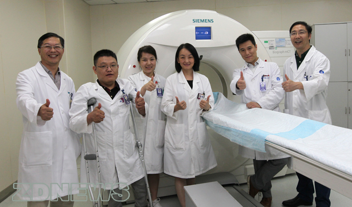
This research involves collaborative efforts by many healthcare professionals in the 2nd Affiliated Hospital of Zhejiang University including Prof. TIAN Mei and Prof. ZHANG Hong from the PET Center, Dr. FENG Jianhua from the Department of Pediatrics, Dr. WANG Shuang and Dr. DING Yao from the Department of Neurology, and ZHU Junming from the Department of Neurological Surgery.
PET detects pairs of gamma rays emitted indirectly by a positron-emitting radionuclide, which is introduced into the body on a biologically active molecule called a radioactive tracer. “Through PET, we can map the whole process of biochemical changes at cellular and molecular levels,” says TIAN Mei.
The success rate of detecting lesions in epileptic children ranges from 15% to 39% according to relevant literature, but PET can elevate the rate to a surprising 79%.
Epilepsy is caused by an abnormal electrical discharge in the brain. This abnormal “short circuit” can cause a person to suddenly have fits and even to lose consciousness.
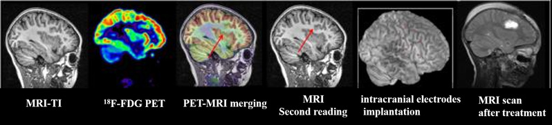
By measuring glucose metabolism with the use of PET, scientists find out that the metabolic rate of the cerebral area with lesions is lower than that of the neighboring area when an epileptic patient is not suffering from any seizure and that this area with no conspicuous energy consumption may well be the seizure area. For some patients, their energy is pronouncedly concentrated, which heralds that a seizure is about to happen. However, this type of energy concentration before the ‘short circuit’ fails to be detected by CT or MRI.
In addition to this, scientists can also pinpoint the location of lesions with the aid of MRI or CT and perform precise surgery accordingly in reference to a database they create. “We figure out a novel diagnostic approach by combining PET-MRI techniques, thus able to develop an effective cure for epilepsy.”
The key to pre-surgery detection is the “positioning system” of PET. By comparing the PET image of a patient with the database, scientists are able to identify the volume of the lesion and keep surgeons well-informed of the area that should be removed via a 3D image.
Because the brain of an epileptic child is far from developed, cognitive damages caused by antiepileptic drugs are exceedingly sensitive. Scientists delve into cognitive impairment—the most common complication of epilepsy.
With the focus on monoamine neurotransmitters, which are involved in a complex neurological system, scientists perform whole-brain imaging regarding the monoamine receptor function and the glucose metabolism of epileptic children. They discover that there appears a significant positive relationship between cognitive performance and monoamine receptor function, thus revealing the mechanism of monoamine neurotransmitters in cognitive impairment. This opens up a potential target for the treatment of cognitive impairment for epileptic children.
Clinical studies indicate that PET molecular imaging technology can not only reflect the changes in cerebral functions and metabolism, but also play an indispensable role in locating lesions precisely, assessing cognitive damages caused by antiepileptic drugs and performing PET-directed surgery, thereby offering a precise plan for the diagnosis and treatment of epilepsy.
“I hope that PET technology, focusing on the idiosyncrasies of different diseases, can be applied to more therapeutic and pre-surgical assessment processes. I also hope that more clinical scientists will get involved in relevant clinical studies, thus providing scientific and appropriate diagnosis and therapy for more patients,” says TIAN Mei.