Raman imaging technique in intraoperative assessment and photothermal ablation of tumor margins
Surface-enhanced Raman scattering (SERS) imaging technique is marked by its superb sensitivity, minimal background interference, and minimal damage to normal tissues for biomedical application. Moreover, the Raman probe has the advantages of high specificity and ultra-low signal interference, thus showing immense promise in the biomedical field. Due to its special application context, most of current researches are primarily focused on in vitro cancer diagnosis, which is confined to the diagnosis and treatment of tumors, such as glioma, in the in vivo state.
Recently, the research team led by ZHOU Min from the Zhejiang University Institute of Translational Medicine made new progress in the precise diagnosis and treatment of abdominal tumors by SERS-based imaging. Their research findings are published in an international peer-reviewed article entitled “Intraoperative Assessment and Photothermal Ablation of the Tumor Margins Using Gold Nanoparticles” in the journal Advanced Science.
Tumors have a high rate of morbidity and mortality worldwide. Abdominal tumors, including orthotopic colon or ovarian tumors, are among the most malignant types of tumors because of their extensive dissemination and metastasis. Since these malignant tumors are highly invasive and metastatic and they can migrate to surrounding tissues and critical organs, surgical resection is clinically challenging, thereby leading to high recurrence and mortality rates.

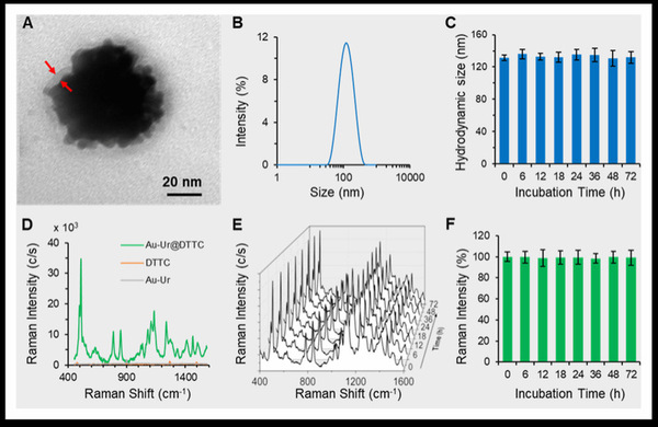
Diethylthiatricarbocyanine iodide (DTTC) is a common infrared dye molecule that has been used as a reporter adsorbed on the surface of the gold nanoparticles in SERS applications. ZHOU Min et al. conjugated the DTTC molecules to the surface of the urchin‐like nanoparticles by using an Au‐S bonding to create an active SERS probe (Au‐Ur@DTTC), wherein the DTTC signal can be used for specific detection of nanoparticles by SERS imaging technique. This probe can be used for intraoperative Raman‐guided resection of tumors. Meanwhile, a post-surgical photothermal therapy (PTT) can also be applied to selective ablation of tumor margin residues, thus significantly reducing the tumor recurrences.

In this study, researchers developed a multifunctional nanoplatform for multimodal bioimaging and treatment, including Raman image‐guided tumor surgery and tumor‐selective PTT. Raman imaging allowed demarcation of tumor margins with high sensitivity and spatial resolution, and SERS mediated pre‐operative mapping and intraoperative imaging facilitated accurate Raman‐guided tumor resection. “Raman imaging, as a high-resolution and sensitive intraoperative approach, shows great promise for targeted detection and ablation of residual tumors after resection within different organs,” ZHOU Min said.