Blood glycoprotain inspires new approach for self-assemly of polymer films
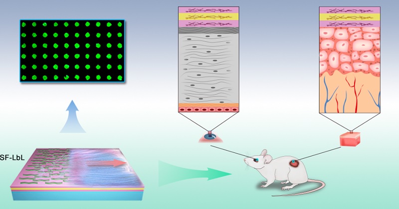
The research team led by WANG Ben, an associate professor in the Zhejiang University Institute of Translational Medicine, develops a shear-flow-driven layer-by-layer (SF-LbL) in situ self-assembly technology. Their research is published in the journal of ACS Nano. This technology not only boosts the assembly speed tremendously and optimizes the surface texture of assembly films but also produces the in situ assembly film on wounded corneas and skin and accelerates the restoration of chronic wounds caused by diabetes.
A counter-intuitive physiological phenomenon can provide inspiration for researchers. It is a relatively formidable task to block a hole in a pipeline in rapidly-flowing currents. However, this seemingly challenging process can be undertaken in blood vessels thanks to a blood glycoprotein termed as von Willebrand factor (VWF), which plays a vital role in the blood-clotting process in high shear stress. At a certain shear rate, this protein converts from a contraction state to a stretch state, exposing enough binding sites to adhere to the surface of the collagen matrix. This allows the formation of a viscous network that further mediates platelet adhesion and blood coagulation. This phenomenon indicates that shear stress has the ability to alter the molecular state effectively.
Inspired by this phenomenon, the research team develops a shear flow-driven LbL (SF-LbL) self-assembly approach that accelerates the adsorption rate of macromolecules by mechanically configuring the polymer chain via a coil-stretch transition, which effectively simplifies and speeds the diffusion-controlled assembly process. The structural characteristics and surface homogeneity of the SF-LbL films are improved, and diverse three-dimensional structures can be achieved. Functional SF-LbL-assembled surfaces for corneal modification are successfully fabricated, and the surface of wounded rat corneas and skin can be directly decorated in situ with SF-LbL nanofilms due to the advantages of this approach. Furthermore, in situ SF-LbL self-assembly has great promise as a simple approach for wound dressing for interventional clinical therapeutics, as is illustrated by the successful in situ fabrication of drug-free layers consisting of chitosan and heparin on the dorsal skin of diabetic mice to rescue defective wound healing.
In theory, any macromolecular substance with electrical properties can be loaded onto the wound surface by this method, so it enjoys extensive applications in materials and drugs. Moreover, it is easy to operate, and it can realize nano-level regulation on film thickness and configuration. The fabricated film is highly adhesive, breathable and transparent. This bioinspired self-assembly approach is expected to provide a robust and versatile platform with which to cure oral ulcer, corneal damage and skin damage.