ZJU team uncovers lysosomal mechanism linking SLC7A11 to Parkinson's risk


A study led by researchers at Zhejiang University School of Medicine has identified SLC7A11, a cystine/glutamate antiporter localized in lysosomal membrane, as an unconventional hydrogen ion (H⁺) transporter critical for maintaining lysosomal pH balance. Published in Cell on April 24, 2025, the work reveals how defects in this pathway lead to toxic protein aggregation and neuronal dysfunction, providing mechanistic insights into Parkinson’s disease and potential therapeutic strategies.
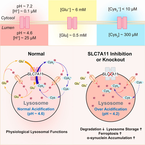
Graphical abstract
Lysosomes, the cell's recycling hub, require precise acidity (pH 4.5-5.0) to fully activate hydrolyses that degrade cellular waste. While the proton pump V-ATPaseimports H⁺, pH homeostasis depends on counterbalancing H+ efflux mechanisms. In 2022, the team discovered TMEM175, a lysosomal H⁺ channel that rapidly releases excess ions during luminal over-acidification. However, when both TMEM175 and V-ATPase were inhibited, lysosomes get unexpectedly alkalized without significant obstacles, suggesting another unidentified H+ “leak” pathway.
“Lysosomal pH regulation resembles a dynamic equilibrium—protons must enter and exit coordinately to sustain enzyme activity,” explained corresponding author Dr. XU Haoxing, Chair Professor at Zhejiang University. “SLC7A11 represents a slow proton release pathway, working in tandem with TMEM175’s rapid response.”
By screening an orphan lysosomal membrane protein (OLMP) knockout cell library, the team pinpointed SLC7A11, a plasma membrane protein based on earlier studies. However, the authors used super-resolution microscopy and biochemical assays to demonstrate its predominant lysosomal localization. Parallel high-throughput screening identified a small-molecule chemical, erastin, which is a widely-used ferroptosis inducer targeting SLC7A11, induced lysosomal over-acidification. Functional studies revealed that lysosomal SLC7A11 exports cystine from lumen in exchange of cytosolic glutamate, whose side chain (pKa ~4.3) undergoes protonation within acidic lysosomes (pH ~ 4.5-5.0). Upon transport to the neutral cytosol, protonated glutamate quickly releases H⁺, creating a continuous H⁺ leak. “Cystine/glutamate exchange across lysosome membrane continuously carries H+from lysosomal lumen to the cytosol through a protonated/hydrogen-dissociated mechanism, so it’s an unconventional lysosomal H+ leak pathway,” said co-corresponding author Dr.HU Meiqin.
“While trying many times, traditional patch-clamp technique failed to detect SLC7A11's activity, likely due to its slow transport kinetics,” said ZHOUNan, co-first author and Ph.D. student. “We have to develop a new assay to evaluate the SLC7A11 activity in single lysosome level, and fortunately we figured it out.”
In the neurons without lysosomal SLC7A11 expression, lysosomal pH dropped below 4.6, compromising the hydrolytic activity of the lysosome, and so accelerating the aggregation of pathological α-synuclein-recapitulated in cells from an early-onset Parkinson's patient harboring a monoallelic loss-of-function (LOF) SLC7A11 mutation. Remarkably, the chloroquine, an FDA-approved drug, in a low-dose to optimally correct the over-acidified lysosome pH caused by SLC7A11 LOF mutation, significantly suppresses the aggregation of pathological α-synuclein.
By integrating SLC7A11's “slow leak” with TMEM175's “rapid exit” and V-ATPase's influx, the researchers revealed a self-complement and self-consistent regulatory framework for lysosomal H+/pH balance. “This closed-loop system ensures the hydrolyses within the lysosome work in an optimal way to clear cellular waste,” Dr. Xu said. “Dysregulation of any component will speed up the pathogenic development of neurodegenerative cascades.”
Dr. XU Haoxing (second from left) with his team
Overall, the findings provide “proof of concept that modulation of lysosomal acidity could represent a therapeutic approach for neurodegenerative diseases,” the researchers wrote.
Source: The research team led by Dr. XU Haoxing